Thermography, iodine tests, hormone tests and vitamin D tests are being touted as alternatives to mammograms in the fight against breast cancer. Established tools like ultrasounds and breast MRIs are also offered up as potential replacements.
You see all kinds of claims on the benefits and dangers of every breast cancer screening tool. So how do you know which cancer detection tool to trust with your breast health?
What's thermography?
While most women have heard of mammograms — it's currently the accepted gold standard for the early screening and detection of breast cancer — thermography is not as widely known. However, thermography has been studied by medical researchers as a potential breast cancer screening tool since the 1970s.
Thermography is an FDA-approved device that uses an infrared camera to produce images of the breast. It picks up the heat and blood flow patterns in your breasts on or near the surface of the body. The use of thermography for breast cancer detection is based on the idea that as some cancers grow, they recruit more blood flow to the area and, therefore, more heat.
When using thermography, there are two main obstacles that reduce its ability to reliably detect early breast cancers. First, we don't often see early, small or preinvasive cancers that have increased the local blood flow enough to be detected this way. Secondly, normal breast tissue is an excellent insulator. So even if there is increased blood flow around a cancer, if a breast cancer is not near the surface of the skin, thermography is unlikely to be able to detect it.
Radiologists want to catch breast cancers as early as possible when they are small and the most treatable. By the time blood flow has increased in the area of a breast cancer to the point where thermography could pick it up, it's often at a more advanced stage of the disease.
Although multiple studies over the last 40 years have shown that thermography is not an effective way to detect and screen for breast cancers, its proponents tout that it has FDA approval. What may not be as widely known, is that thermography received FDA approval many years ago when only safety had to be proven, not efficacy. So thermography as a technology has been shown not to have any direct harm to the breast tissue, but it has also not proven itself to do what it says it does — reliably detect breast cancer.
In fact, thermography is not approved as a stand-alone tool for breast cancer screening. The FDA has regularly warned thermography facilities about marketing and using the device as a standalone breast cancer tool because there is no medical evidence that shows that thermography, used alone or along with mammography, is an effective screening for breast cancer.
What makes mammography a better option?
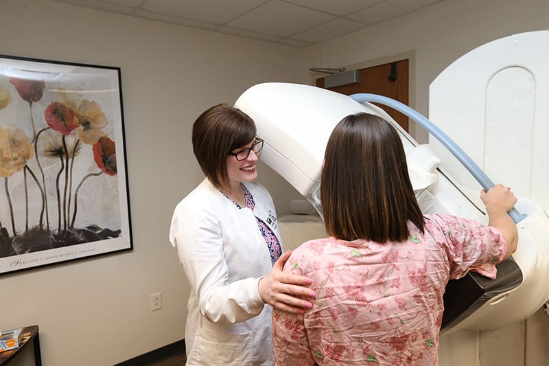
The CDC, American College of Radiology, Society of Breast Imaging and all the other major professional organizations involved in breast health agree: mammograms are the best way to find breast cancer early. Here are the facts:
1. Annual screening mammograms save lives.
This is no claim. It's backed by extensive research. As many as 13,000 women are saved every year from breast cancer death as a result of screening mammography. According to the American Cancer Society, screening mammograms have reduced the death rate from breast cancer by 39 percent over the past 30 years.
2. False positives and overdiagnosis are rare.
False positives happen in any screening test. They're not unique to mammography. In screening mammography, an exam is considered a false positive if you're called back for additional pictures after your screening study to ultimately find out if the area of concern on the screening exam is a normal finding.
Although getting called back in for further testing can be worrisome, it doesn't happen that often. Only 1 in 10 women get called back for additional diagnostic testing — a rate similar to other cancer screenings like Pap smears. Of those 10 percent, over half ultimately find out that they're in the clear.
Recently, some have raised concerns of overdiagnosis in screening programs, meaning that a cancer is present and detected but it may be so small and slow growing that your overall health and longevity wouldn't have been affected had the cancer not been detected and treated.
The idea of overdiagnosis applies to a very small number of focal pre-invasive cancers, or DCIS. The natural history for many pre-invasive cancers if left untreated is to progress to invasive cancer and then ultimately metastatic disease. While there are some cases of DCIS that may progress slowly enough for watchful waiting to be appropriate — especially in patients who may be poor surgical candidates due to other health problems — we currently have no way to tell the fast-moving, aggressive cases apart from the slow-growing cases.
Without a way to distinguish between which pre-invasive breast cancers will progress rapidly, and which will move slowly, breast specialists are obligated to treat every cancer as if it could progress. The concept of overdiagnosis is really more of an intellectual debate than anything at this point.
3. Adding 3D imaging to your mammogram catches more cancers and decreases callbacks.
3D mammograms can offer a better, more well-rounded visualization of the whole breast by giving the radiologist multiple thin-slice images to look at in each view instead of only one image of all the breast tissue. This makes it easier to find small cancers that might otherwise be hidden by normal breast tissue.
In fact, some studies show that the cancer detection rate for small, invasive cancers increases by up to 40 percent when 3D imaging is added. You are also less likely to get called back from a screening for normal overlapping tissues because the 3D images can often separate them.
While ultrasounds and MRIs are also used to produce additional images and look at the breast tissue in different ways, these tests are not recommended by the American College of Radiology as the first line test for screening average risk women. There are certain early signs of breast cancer such as calcifications and distortions that are best seen on a mammogram. Ultrasound and MRI can be important in certain settings, but they do not replace annual screening mammography.
When should I schedule my first mammogram?
If you're a woman over the age of 40, right away! Every woman should get an annual screening mammogram starting at age 40 — just as you get a Pap smear from your OB/GYN to check for cervical cancer. No physician referral is required for women over the age of 40.
You shouldn't wait until age 40 and your first mammogram to worry about your breast health though. The American College of Radiology recommends that every woman have a risk assessment before age 30 to see if regular breast cancer screening or supplemental screening is needed earlier than age 40. But if you have a problem or concern about your breast health at any age, such as new lumps, skin changes, persistent pain or nipple changes, you should see your primary care provider right away for a clinical evaluation and possibly diagnostic imaging of the breasts.
When it comes to early breast cancer detection, there are no substitutes. The medical community is in agreement on it. Whether it's your first mammogram, it's been a while or you just need a risk assessment, schedule an appointment to start managing your breast health — it could save your life.
